HYPEROPIA (Long-sightedness)
Hyperopia can cause difficulties in focusing at near, such as reading and computer work. If there is significant hyperopia, it can also blur distant objects as well.
With this extra focussing, one can experience eye-strain, headaches and blurred vision especially after prolonged close work.
The eye works by focusing an image clearly on the retina at the back of the eye so as to attain clear vision. With hyperopia the image is focussed behind the back of the eye and will appeared blurred. However, the focusing muscles inside the eye will attempt to clear the image by focussing the image on the retina.
The solution is usually glasses to relieve the eye-strain and headaches.
 MYOPIA (Short-sightedness)
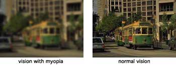
Myopia causes blurred vision for distant objects. It can be difficult to read road signs and scoreboards. It can also be difficult to recognise people in the distance.
The eye works by focusing an image clearly on the retina at the back of the eye so as to attain clear vision. With myopia the image is focussed before it reaches the retina and will appeared blurred.
Myopia tends to begin in teenage years and may worsen as the eye grows to full adult size.
Causes of myopia are not known for sure. There are various theories including hereditary factors and environmental factors such as poor diet and excessive amounts of reading and close work.
Myopia can be corrected with either contact lenses or glasses.
ASTIGMATISM
Astigmatism occurs when the eye is not perfectly round. An eye with astigmatism is not perfectly round but is shaped a little like a football. It is not a disease as it relates purely to the shape of the eye. Because of the irregular shape, the eye cannot focus the image clearly on the retina at the back of the eye, resulting in blurred vision.
Astigmatism can cause eye strain and discomfort and possibly headache after concentrating on an object. For more severe astigmatism the vision may be blurred at all distances.
Spectacles and contact lenses can be used to correct astigmatism.
 TEST FOR ASTIGMATISM
Close one eye and then the other one, if you do not see all the lined squares, in the same black colour, or if you see one or more squares greyer than the others, you than have astigmatism. Consult your optometrist to examine your eyes to determine the amount of astigmatism that you have, and if it is affecting your lifestyle.
 PRESBYOPIA (Over 40 vision)
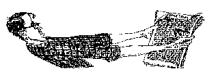
This is a gradual loss of focusing ability of the eye. It is a natural change that happens to all of us. It becomes more and more difficult to read and thread needles. It seems like our arms are not long enough, as we tend to push the book further away from us to see clearly.
Presbyopia is usually first noticed around the age of 40 to 45 years.
A normal hardening of the lens inside the eye causes presbyopia. As the lens loses flexibility it is less able to change shape and hence close focusing becomes difficult.
Presbyopia is corrected by spectacles for reading or can be in the form of bifocal spectacles or multifocal spectacles. It can also be corrected with multifocal contact lenses.
EYE DISEASES
GLAUCOMA
Glaucoma is a disease in which the nerve cells, which transmit information to the brain, become damaged. In most people this damage is due to an increased pressure within the eye. In other people the damage may be due to poor blood supply to optic nerve fibres at the back of the eye, a weakness in the structure of the nerve or a problem with the health of the optic nerve itself.
Over 300,000 Australians have glaucoma. It can affect people of any age, but is more common in older people.
People with a family history of glaucoma are more at risk and should have their eyes examined regularly.
There are usually no symptoms with glaucoma until permanent damage to the eye has occurred. If left untreated glaucoma may result in blindness.
The best way to detect glaucoma early is to have regular eye examinations. At these examinations, the pressure of the eye will be measured; the optic nerve will be examined. If needed a visual field assessment will be performed using a computerised instrument that maps out your side vision.
All these tests are simple and painless and are performed at our practice. If we detect glaucoma, you will be referred to a glaucoma specialist to manage the condition.
Glaucoma is usually treated with eye drops or surgery. Glaucoma cannot be cured but can usually be controlled to prevent or slow down further loss of sight.
 CATARACT
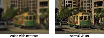
Cataract is a clouding of the lens inside the eye which causes blurring of vision. It is not a growth and cannot be seen on the surface of the eye.
It is like looking through a dirty windscreen, vision is not as clear as looking through a clean windscreen. With a cataract, vision appears hazy or blurred.
Cataracts are commonly a result of ageing and long-term exposure to ultraviolet light. They can also be caused by injury and disease. Occasionally cataracts are present at birth.
As cataracts progress the patient's sight worsens. This can take months to years to occur. Cataracts usually affect both eyes, although they can progress at different rates. It is common to see cataracts in people over 65 years.
Treatment is usually needed when your vision interferes with your life style. Sometimes changing your spectacle lenses can improve your vision. Once the cataract is fairly advanced spectacles will not clear your vision and surgical treatment, which consists of surgically removing the cloudy lens and replacing it with a clear artificial lens (intra-ocular lens implant) is needed. After surgery your vision should be excellent.
DIABETES
People with diabetes can develop eye problems such as glaucoma,cataracts and double vision. The main sight threatening part of diabetes is its effect on the retina.
After diabetes has been present for some years, changes may occur at the retina which lines the back of the eye and is responsible for transmitting light messages to the brain vis the optic nerve. These changes are known as diabetic retinopathy.
Diabetic retinopathy causes changes in the small blood vessels in the retina, leading to weakening and leaking, which forms small haemorrhages. This can cause swelling of the retina. If not treated permanent damage to the retina can occur and lead to loss of vision.
The risk of developing diabetic retinopathy increases with the length of time the patient has had the disease. The risk also increases with poor control of blood sugar levels.
At our practice all diabetic patients are carefully examined after drops are placed on the eyes to enlarge the pupils. This allows for a detailed examination of the eyes.
It is recommended that people with diabetes have their eyes examined every year.
If we find any sight threatening signs we will refer you to a retinal specialist to manage the condition.
 AGE RELATED VISION LOSS
Some people lose their central vision as they age. This can be a gradual or sudden loss. People with this problem still have side vision but have difficulty seeing straight ahead.
This is a condition affecting the macula, the central part of the retina at the back of the eye. It is known as Age Related Macula Degeneration. The macula has the highest density of light sensitive nerve cells and is the part of the retina we use for sharpest vision.
Macula degeneration occurs when nerve cells are damaged or deteriorate. When this happens, your central vision is significantly affected.
Some degeneration leads to leakage from the tiny blood vessels in the retina. This can lead to swelling and haemorrhages. This type of macula degeneration can be helped with laser treatment to the retina.
Unfortunately a lot of macula degeneration cannot be treated.
This condition mainly affects older people, usually over 60 years. It appears to be more common in smokers and run in families.
At our practice we examine peoples eyes after using eye-drops to enlarge their pupils. This allows us to closely examine all the structures of the eye. If leakage of blood vessels is observed the patient is referred to a retinal specialist for treatment.
PTERYGIUM (pronounced te-ri-gi-um)
A pterygium is a triangular-shaped lump of tissue on the white of the eye, usually on the side closer to the nose. It can sometimes grow on to the clear part of the eye known as the cornea.
A pterygium results from long-term exposure to ultra-violet light, wind and dust.
If the pterygium grows across the cornea it can distort the vision.
Treatment of the pterygium can be sunglasses to protect from ultra-violet light and dust. Sometimes lubricating eye-drops can soothe the irritation. If severe, surgical removal may be necessary.
Patients with a pterygium should always wear glasses with a UV filter when they go outside.
SPOTS AND FLOATERS
Some people notice a 'hair or spot' in their vision. It can be quite annoying as you try to brush it away, but to no avail.
Spots and floaters are usually semi opaque specks or particles of protein within the eye that are sometimes seen in the field of vision. These particles are suspended in the fluid within the eye. They can appear as strands, dust-like particles or cobwebs. Because floaters move as the eye moves, they jump away as you try to look at them.
Cells left over during the development of the eye before birth can cause floaters. They can also be caused by injury or eye disease.
We recommend a complete eye examination to determine the cause of the floaters.
KERATOCONUS (Cone shaped cornea)
Keratoconus is a thinning of the central zone of the cornea, the clear front surface of the eye. As the cornea thins it bulges forward in a "cone' shape, causing distortion and blurring of vision.
Keratoconus usually occurs between 10 and 25 years of age and is an inherited disorder.
Treatment consists of glasses initially. If the condition progresses special rigid gas permeable contact lenses may be needed to give clear vision again. In severe cases where contact lenses cannot be fitted to correct the vision, a corneal transplant is needed.
We have a lot of experience in treating people with keratoconus with contact lenses. It is important that people with this condition have regular eye examinations to assess the fit of the contact lenses. If the cornea changes shape the contact lens may need to be changed to fit the eye correctly.


